News
What the human heart looks like from the inside: scientists have shown the difference between a healthy and a sick organ. Impressive video

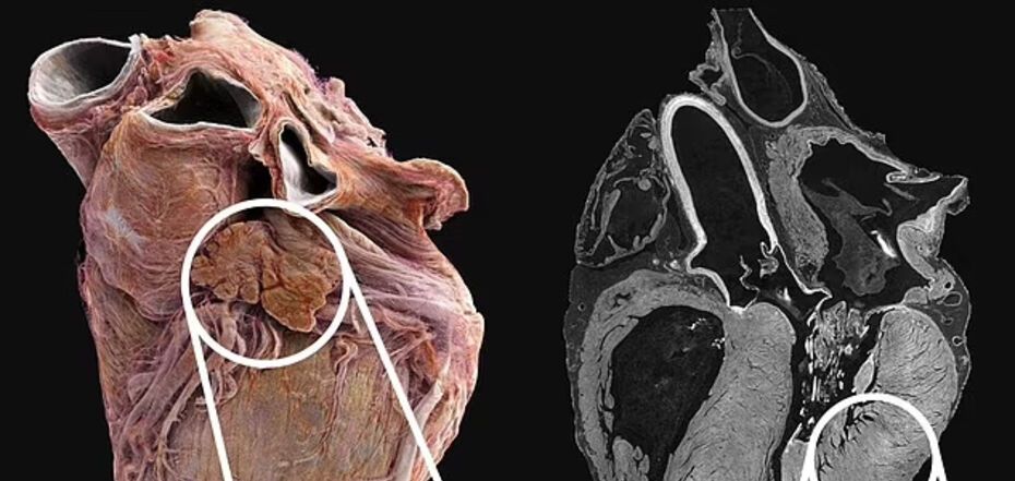
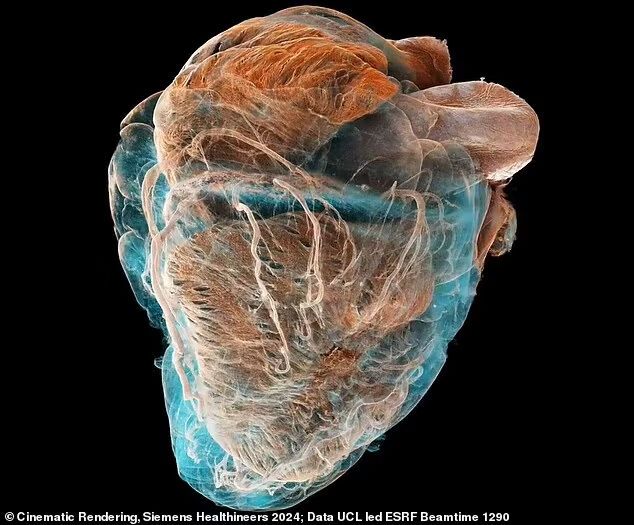
Comparison of sick and healthy hearts.


With the help of a video, scientists have shown how the human heart looks like from the inside. It is possible to see the difference between a healthy organ and the organ of a patient with cardiovascular disease.
Specialists from Britain and France used a new X-ray technique that captures the structure of the heart with an accuracy of 20 micrometers (half the width of a human hair). In some areas, visualization is done at the cellular level, that is, individual cells of the organ are shown, writes DailyMail.
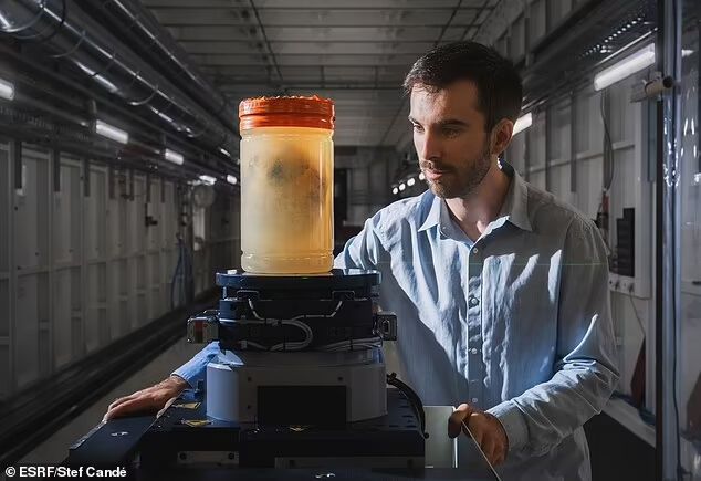
Experts from University College London (UCL) and the European Synchrotron Radiation Facility (ESRF) in Grenoble (France) emphasized that the new technique opens up new possibilities for the study of human organs. "It allows you to see the whole organ on a global scale and then zoom down to street level to look at features of the cardiovascular system in unprecedented detail," said Peter Lee, Professor of Mechanical Engineering at UCL.
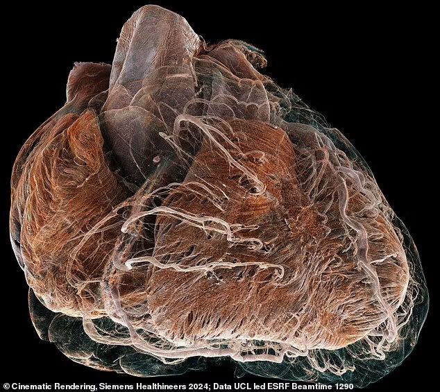
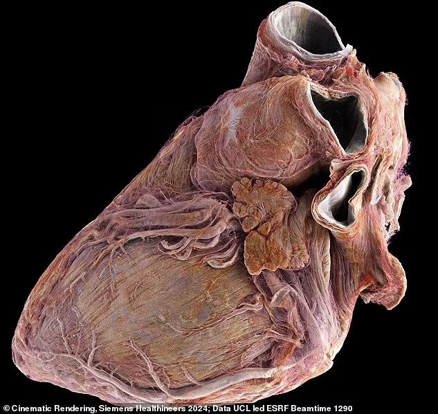
The video compares two whole organs from deceased adult donors. The healthy heart is from a 63-year-old man. The diseased one is from an 87-year-old woman with a history of coronary heart disease, when the organ is weakened due to decreased blood flow. The difference is obvious: a healthy heart has a well-defined shape, while a sick heart is rounder, with dried vessels and muscle fibers.
A team of scientists used an X-ray technique called hierarchical phase-contrast tomography (HiP-CT) to image the heart at a scale of 20 micrometers.
"One of the main advantages of this technique is that it provides a full 3D image of the organ, which is about 25 times better than a clinical CT scanner. In addition, the new technology can zoom in to the level of cells in selected areas, which is 250 times better to achieve the same detail as under a microscope, but without cutting the specimen.
However, it is not possible to examine the heart of a living person in this way because the radiation dose would be too high.
Experts believe that these images will be a source of better understanding of cardiovascular disease. After all, according to the Ministry of Health, coronary heart disease was responsible for 8.9 million or 16% of deaths worldwide in 2019. And that number has increased by more than two million since 2001.
Only verified information from us in Telegram-channel OBOZ.UA and Viber . Do not fall for fakes!
Other News


The long-awaited VW crossover cheaper than Duster was shown in photos
The Tera is being prepared for the premiere

How to boil beets in 10 minutes: an elementary method that will suit everyone
You don't even have to turn on the stove


Delicious and healthy "Napoleon" made from phyllo dough: it takes a few minutes to prepare
The easiest way to make a popular dessert

The most fashionable manicure color for spring 2025: five periwinkle designs
Spring 2025 brings a new color favorite in the world of manicure